
NOTE:Most hyperlinks in this resource are to external web pages that will appear in new browser windows. To return, please just close the window containing the external resource.
Occasionally I've condensed the text. Click "more . ." to expand or close the hidden descriptions. Any comments or other feedback may be sent to me via LinkedIn or Research Gate (links at the bottom of this page). I hope you will enjoy browsing this resource. Michael Geisow 2026.
You can select full screen mode [ ] or the speed of playback and other functions can be set by clicking on the 3 vertical dots
Referring to the AnxA5 structure above, the annexin "core" takes the form of a saucer-shaped "washer" with a convex surface (top of image above) which carries the binding sites for Ca2+ ions and membrane lipid head groups. At the centre of this core is a hydrophilic channel (image right). The core is made up of 5 homologous α -helical bundles (coloured separately in the image) making up structural domains each carrying 2 Ca2+- and lipid- binding motifs. These motifs have been termed type II calcium binding structures to distinguish them from the more common type I calcium binding structures in parvalbumin and the S100 protein family. Although these structural motifs are homologous, both Ca2+ and lipid binding affinities differ between annexins: some annexins do not display Ca2+-dependent membrane binding, but associate with membranes by other means.
The lower convex protein surface carries the C-terminus and the N-terminus which is different in length and sequence for each annexin. In the illustration above this text the chain termini can be distinguished in the lower left. The N-termini are dynamic in most annexins complexed with Ca2+ ions and in plant annexins are reduced in length compared with animal annexins [these N-termini have also traditionally been called "domains" in the annexin literature]. The concave annexin surface forms the binding site for partner proteins such as members of the S100 Ca2+ binding protein family. In fact, most of the molecular surface of annexins appears to interact with partner molecules as will be described later. You can animate and interact with the pig AnxA1 (illustration right) by clicking on the static image shown! The molecular representation displayed can be rotated and zoomed and interrogated (click within the circle top right) to display detailed residue conformations and interactions.
Currently, this canonical annexin core structure appears to be conserved in almost all eukaryotic organisms studied. This is testament to the significance and antiquity of the annexin protein fold. However, in the diverse kingdoms which constitute the eukaryotic domain of life, annexin orthologs (homologous proteins in different species) now serve and can further be expected to serve a wide range of different functions. The presence in most single species of multiple paralogs (homologous proteins in the same species arising from gene duplication events), further indicates the value of the annexin family to virtually all forms of eukaryotic life on earth.


Annexins are almost universal eukaryotic proteins and analogous proteins may even be present in some prokaryotes. Almost all organisms; multi- or unicellular, possess multiple annexin paralogs There are up to 25 annexin paralogs in bread wheat; all with very similar core sequences, but non-homologous amino terminal sequences. Humans express 12 annexin paralogs. So far the sequenced annexin transcribed genes (there are pseudogenes) code for either 1 or 2 of the homologous tertiary structural cores described above. [NOTE: It has become traditional in the annexin literature to refer to these cores as domains, whereas (in my opinion!) the term domain should more accurately refer to the 4 homologous repeated 5-helix bundles which make up the annexin cores.] Annexin amino terminal sequences are highly variable in sequence and length, structurally dynamic and considered major contributors to the diverse functions of this protein family. It is notable that analogous dynamic protein regions (e.g. unstructured in x-ray crystallographic studies) participate in binding with other protein partners - when they adopt defined secondary structure - often α -helix in the case of annexins.
Those annexins studied in detail have been shown to be under numerous transcriptional and post-transcriptional controls. Regulation by micro (miRNA) and other non-coding RNAs is an increasing area of study; especially in tumorigenesis. Annexin functions are further subject to post-translational modifications (PTMs), most prominently by phosphorylation at serine, threonine and tyrosine residues. Further PTMs identified include sumitoylation and fatty acid acylation: especially by palmitic and myristic acids


Annexins are "Janus" proteins: found free or facing outwards in association with extracellular membrane surfaces as well as located within or facing inwards in association with plasma and organelle membranes towards the cell cytosol. They are not intrinsic membrane proteins, but associate either dynamically or constitutively with plasma and organelle membranes and participate in membrane trafficking processes, sculpting at specialised membrane regions through linkage to cell cytoskeleton and modulating membrane lipid composition. Found in most organisms both in cytosol and in association with cell surfaces, they appear to participate in cell and tissue homeostasis; for example in the resealing of damaged plasma membrane. Their presence and functions both within the cytosol as well as the extracellular space may be a distribution almost unique to these proteins, but is logical to the extent that they interact with lipids which are present in both halves of membrane bilayers
Annexins have a wide range of functions, but generally appear to participate in both sensing and responding to stress: abiotic and biotic in plants (pH, salinity, osmotic pressure, membrane damage, pathogens) or biotic in the animal kingdom (infection, vascular pathologies). In animals at least they are coming to be considered important players in innate immune systems and in humans, important players in the inflammation that follows traumas. This appears to be a double edged sword: Their presence on cell surfaces, especially vascular endothelia, leads to the opportunistic adhesion of specific bacteria and viruses: a prelude to infection. Dysfunctional regulation of the production of certain annexins (hypo- or hyper-expression) is now seen as a risk factor in pathogen infections and an indicator of tumorigenesis
Annexins are intimately involved in signal transduction and are broadly implicated in paracrine, autocrine and even endocrine pathways. Intracellularly they act as adaptors, molecular scaffolds, transducers and effectors and, in this way are involved in functions such as apoptosis, mitosis, cell proliferation, migration and tumorigenesis! In their varied amino termini and their PTM, they expose recognition sequences for partner protein SN2 domains (which recognise phosphotyrosine) and SN3 domains (which recognise polyproline regions). This together with their recognition of Ca2+ ions and lipids like PIP2 means they can act as intermediaries in many different signalling pathways.
A considerable amount of data has accumulated concerning the altered expression or location of annexins in pathologies. The well studied anticoagulatory and anti-inflammatory effects of certain annexins (especially AnxA1 and AnxA2) have suggested that these annexins, in recombinant or biomimetic form, could be valuable in therapeutic interventions. This aspect is reviewed in depth in the "Annexins in Medicine tab". It is expected that this section of this resource will see the most frequent updates!
For such a large, relatively abundant and pan-eukaryotic protein family, it has taken a remarkable amount of time for explanations of function to emerge. And that seems to be because they have multiple functions, even when considering a single annexin (vertebrate AnxA2 is a good example here). The evolutionary history and rationale for annexins is intriguing and yet to be explained. Also mysterious for such a universal protein family is the outcome of many annexin gene knock out studies. Removal of one and sometimes several annexins does not appear to greatly affect normal cell, tissue, even whole animal growth and development. For many similarly universal intracellular proteins, loss or mutation might be expected to prove catastrophic. It is agreed, however, that annexins are called into play when cells and tissues are subject to stresses, in both animal and plant kingdoms at least.
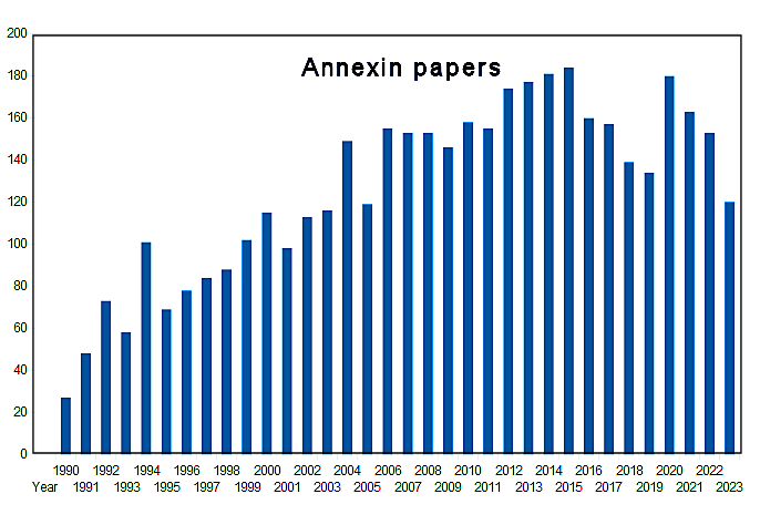
The annexins form a fascinating and enigmatic family and are making their presence felt now in areas as different as medicine and agriculture as this website hopes to demonstrate/ This conserved protein family has turned out to have pan-eukaryotic orthologs (homologous proteins arising through speciation) following the widespread genome programmes. And the nomenclature has been extended: Across the biological domain eukaryote, sequencing has so far identified between 1 and 29 annexin gene paralogs in vertebrate (Ana) (Moss and Morgan, 2004), invertebrate (Nab) (Cantacessi et al., 2013), fungus (AnxC) (Khalaj et al., 2015), plant (AnxD) (Jami et al., 2012; Wu et al., 2022) and protist (AnxE) (Einarsson et al., 2016) kingdoms. Bioinformatic surveys of bacterial genomes have identified a possible limited number of annexin gene orthologs (AnxF) (Kodavali et al., 2014)
In many ways, free living single cell organisms provide easier models for mechanistic research. But even here, annexin research has been perplexing. Fission and budding yeasts have plasma membranes with lipid compositions similar to most other eukaryotes. Hence lipid transport is comparable to that in eukaryotes. These yeasts possess the full complement of eukaryotic organelles, have membrane trafficking, cytoskeleton proteins and efficient endo- and exocytotic pathways, the latter even perfectly capable of transcribing, processing and secreting heterologous human proteins. Their numerous intracellular signalling pathways include G-protein linked receptors, multiple protein kinases, cAMP and Ca2+ -signal transduction (Schultz et al. 1995), yet they lack genes for annexins (Khalaj et al., 2015). For these eukaryotes at least, annexins are dispensable. It may be significant that yeasts have exceptionally tough and cross-linked glycan cell walls surrounding their conventional lipid membranes. In addition to this protective physical barrier they express other proteins, unrelated to annexins under conditions of biological and physiochemical stress. But these yeasts do serve to emphasise the fact that eukaryotic life can go on without annexins. Or perhaps it would be more correct to suggest that in the case of certain yeasts, other gene products serve roles that annexins play in most other organisms.
Future directions New approaches to understanding functions and certainly incorporating the annexin family in diagnostic and therapeutic medicine will require development of new model systems such as tissue explants and organoids. Multi-omics approaches (proteomics, transcriptomics, epigenomics etc.) will need to be employed. Studies of the epigenetic control of annexin expression are presently in their infancy, but such studies - especially applied to development and pathologies - will surely increase. There are already indications of this: (Integrative Modelling Reveals Annexin A2-mediated Epigenetic Control of Mesenchymal Glioblastoma Kling et al.).
NOTE: This section reflects on the early recognition of the Ca2+-regulated proteins later to be known as the annexin family. It refers to work carried out in the period 1975 - 1990. The content is based around my own studies, but many international groups were independently working with and naming these proteins. Since 1990, the annexin field has exploded and references can be found in the other sections of this web resource.

Analogous proteins were reported from animal tissue extracts from around the late 1970s. The researchers in the groups responsible were all studying different biological activities and the only then apparent connection between the different proteins reported were their calcium binding properties. This in itself was not especially noteworthy as cells express many different calcium binding proteins. Two labs were interested in the molecular mechanism of triggered exocytosis: our own lab and that of Harvey Pollard in the USA. Secretion was known to be driven (or at least dependent) upon an increase in intracellular Ca2+ concentration. Baker and Knight in the UK showed that secretion of adrenaline from electro-permeabilised adrenal chromaffin cells could be triggered by raised extracellular Ca2+ levels. My lab (at the National Institute for Medical Research outside London) and Carl Creutz in Pollard's lab at the University of Virginia, decided to use the isolated secretion granules to investigate potential trigger proteins either on the granule membranes or in the chromaffin cell cytosol. Creutz was the first to report a cytosolic protein, which he named synexin, (now known as AnxA7) which aggregated chromaffin granule membranes in a Ca2+ - dependent manner.


Later, we reported the isolation of a group of calcium-dependent chromaffin granule membrane binding proteins (see Geisow & Burgoyne (1982), which at that time we described simply by their apparent molecular weights on dissociating 1D and 2D SDS gel electrophoresis: P70 (AnxA6); p32 (AnxA4) p35 & p35.5 (AnxA1, AnxA3 & AnxA5) & p36 (AnxA2). At first we did not see Carl Creutz's 47kDa synexin (AnxA7) possibly because we had used lower free Ca2+ levels in our experiments. The 2D separation is illustrated on the left.
At that time we assumed that all these proteins arose from chromaffin cells, but of course such dissected tissue extracts contain many other cell types e.g. vascular leucocytes and other epithelial cells which would have contributed lower levels of annexins. It must also be recognised that the cattle adrenal glands would have been subject to substantial stress for some time during transport. It is more than likely that blood levels of adrenaline and cortisol would be high for some time and would have exerted endocrine and paracrine effects, possibly even de novo annexin synthesis (and as we now know), extracellular translocation of some of these annexins. When we searched the literature it was evident that a number of labs had isolated proteins of very similar molecular weights (32KDa - 36KDa and 70KDa) which bound reversibly to membranes isolated from a range of tissue types; not just secretory organelles. I compared our adrenal proteins with some of those from other tissues by 2D gel electrophoresis and found identities or close similarities.
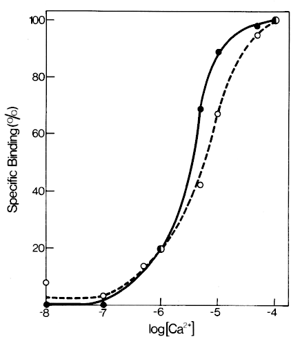
Radio iodinated Ca2+ dependent binding proteins bound to resealed granule membranes in a microfuge sedimentation assay. (Geisow & Burgoyne (1982)). Although at that time the assay used a mixture of proteins, the highest radio labelling was of the 70kDa protein (AnxA6) - probably because of the greater tyrosine content. Half maximal granule membrane binding occurred at 2 μM approx.
We also tried radiolabelled calmodulin, which did bind with membranes, but was not seen in our recoveries of adrenal medulla cytosol proteins and the stoichiometry of calmodulin / granule membrane binding (wt/wt) was less than 0.005%. We did not investigate calmodulin binding to granule membranes further at that time. Treatment of resealed granule membranes with proteases did not abolish the 32kDa- 36kDa and 70kDa Ca2+ dependent binding, but did reduce calmodulin binding.

Initially we used whole chromaffin granules to isolate annexins. Subsequently we found that annexins bound just as well to granule membrane "ghosts" and even membrane ghosts treated with proteases. We noted it was possible to purify annexins in a single Ca2+ - dependent affinity purification step on phenyl Sepharose. Membrane ghosts were trapped by hydrophobic interactions with the affinity column allowing facile isolation of annexins even from whole tissue acetone extracts. The figure shows that it is the acidic phospholipids which were specifically recognised by annexins. Later others - especially Volker Gerke - showed that annexins combine with polyphosphoinositol lipids.
The blue arrows in the 2D gel image shown earlier indicate particularly strong cross reaction with spots representing AnxA2, AnxA4 and AnxA6. Several of these proteins were later found to to be substrates for protein kinases and phosphatases. In particular, the clear horizontal splitting of the spot AnxA2 in the pI (isoelectric) gel axis (see 2D gel image) may be due to phosphorylated and dephospho - forms. Spot Anx2 was later shown to be a substrate for both serine and tyrosine kinases. A number of the other proteins in the gel image (AnxA6 for example) also showed this splitting.
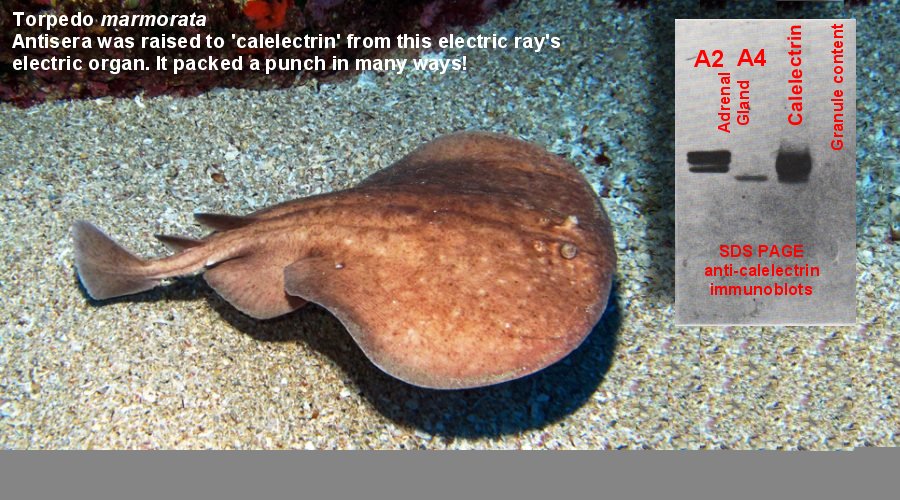
At that time I was fortunate to have been setting up a highly sensitive gas phase protein sequencer and began to sequence tryptic peptides from our adrenal proteins and calelectrin. We were in for a surprise: one that went some way in explaining the curious cross-reactivity of our anti-calelectrin immunoglobulin. The close annexin sequence homologies and the fact that the antiserum had been raised against denatured calelectrin may have explained the relatively broad specificity of ray anticalelectrin towards our bovine proteins.

All the annexins we sequenced contained up to 4 highly homologous 17 amino-acid repeats. We knew Michael Crumpton's lab at the Imperial Cancer Research Fund (ICRF) in London were working on an analogous Ca2+-binding protein from lymphocytes and in a rather tense meeting we exchanged some unpublished sequence information. Michael Crumpton had been in a long queue for access to the only other UK gas phase protein sequencer in Mike Waterfield's lab at ICRF, but finally had some peptide sequence information. A sequence clearly homologous with those in our proteins was also present in Mike Crumpton's 70kDa lymphocyte protein (later identified as Anx A6). Wasting no time on further sequencing, we submitted a letter to Nature which was accepted (Geisow et al. 1986). We did not know at that time that the cDNA of one of these proteins – lipocortin 1 (AnxA1) had been cloned by the US biotech firm BIOGEN and its full sequence was published in Nature shortly after the acceptance of our own paper. Remarkably, the authors from BIOGEN did not point out the clear internal 4-fold homologous repeats present in their published sequence! We soon suspected that these repeats were part of the Ca2+ and lipid binding sites common to all these proteins. I have aligned some of our experimentally-determined peptide sequences against those obtained from cDNA sequencing (shown in the figure below).


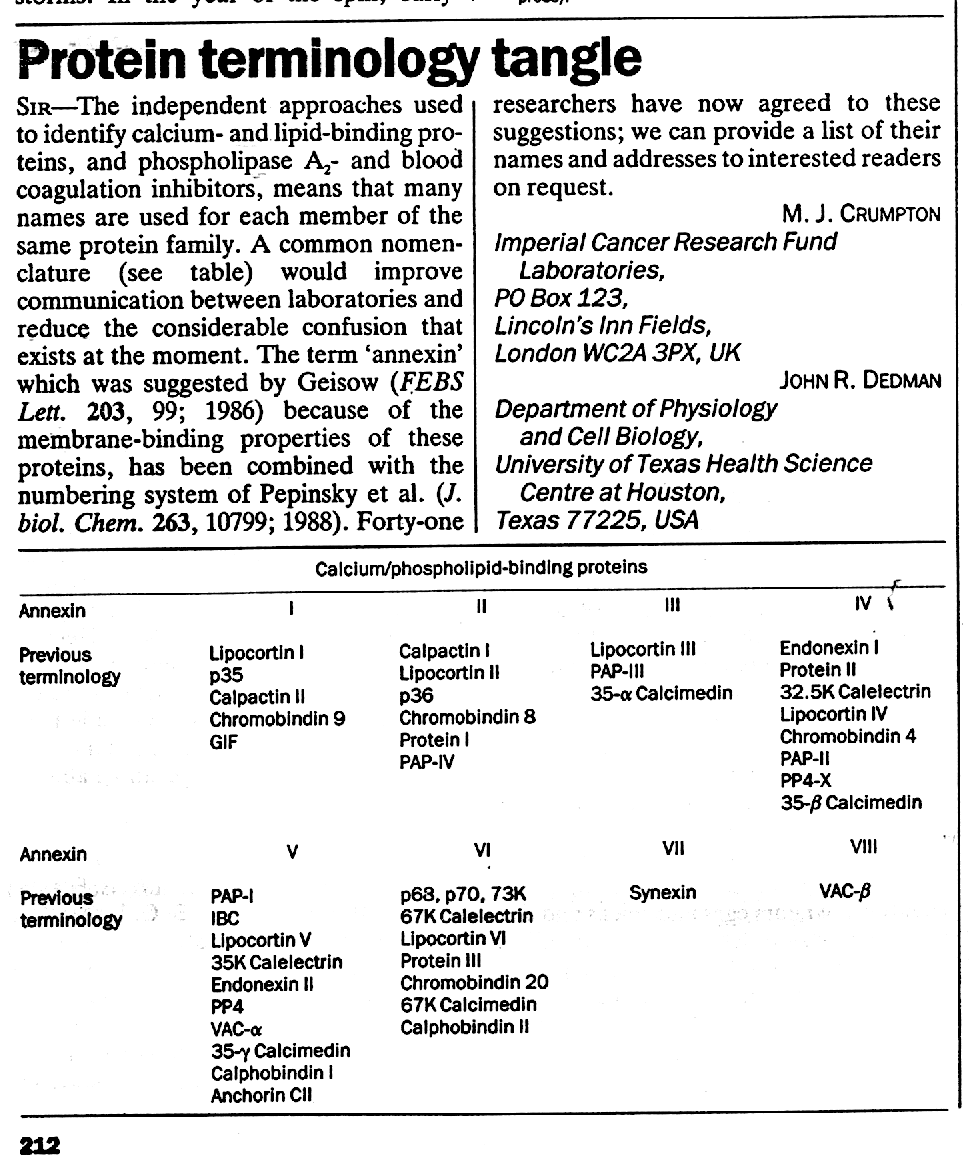
At the time of our and Carl Creutz's discovery of the annexins in adrenal gland, the number of reports and different names of analogous proteins grew: Lipocortins: anti-inflammatory factor(s) in blood plasma; Chromobindins: multiple Ca2+-dependent chromaffin granule membrane binding proteins; VACs: Vascular AntiCoagulants α and β Calpactins: with anti-phospholipase C activity; PAPs: Placental Anticoagulant Proteins; Calelectrins: Ca2+-regulated proteins from the electroplax glands of electric ray; Calphobindins: anticoagulant and protein kinase C inhibitory proteins from human placenta; Anchorins: surface collagen binding protein(s) identified on chondrocytes; PP4s: Placental Protein 4 inhibitors of phospholipase A2 and thromboplastin; Calcimedins (initially from chicken gizzard) with phospholipase A2 inhibitory activity. I had determined most peptide sequences on AnxA4 which I was then calling endonexin, inspired by Creutz's name synexin for AnxA7. One of the most striking aspects of annexins is the structural motif containing the Ca2+ -binding site. Many papers still refer to this motif as the endonexin fold; for example in the recent sequence mutation work of Harvey Pollard's lab, where synexin was first reported.
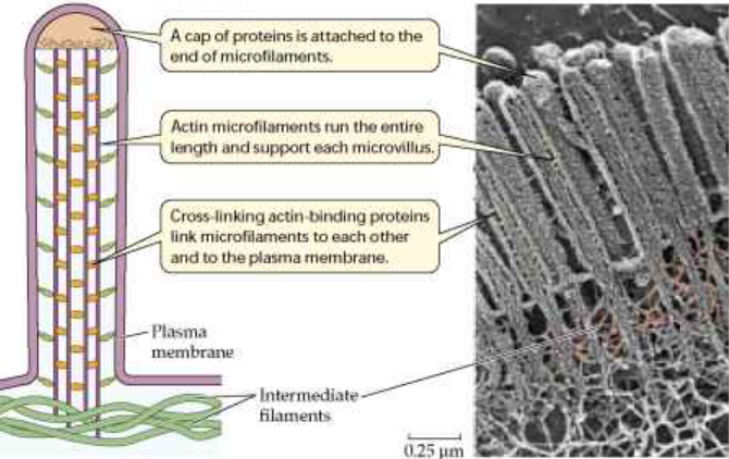
We had noted and published on the phosphorylation of chromaffin cell proteins exposed to secretagogues. We obtained a specific antiserum for pp36 - a major substrate for the Rous sarcoma virus src-gene tyrosine kinase- a kind gift of Sara Courtniege - and found that our p36 (Anx A2) protein was immunoprecipitated. Volker Gerke in Klaus Weber's lab in Munich, Germany had been intensively studying a 36KDa protein present in intestine (localised in the brush border membranes of epithelial cell microvilli). He had independently reported that this protein (now known as AnxA2) was phosphorylated by Rous sarcoma virus tyrosine kinase. He was also able to show that AnxA2 combined with Filamentous actin (F-actin) and that it formed a non-covalent heterotetramer with two molecules of an S100 protein. Since those early days, Volker has become one of the leading researchers on mammalian annexins.

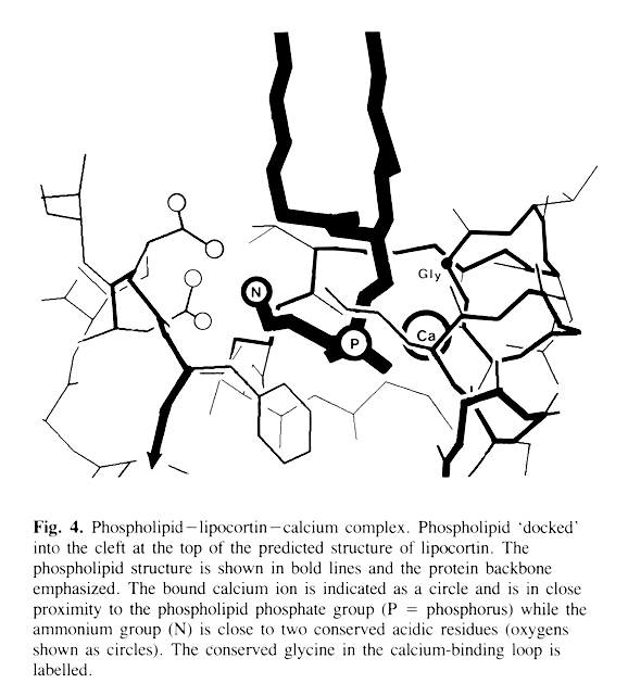
Excited by our discovery, I contacted Willie Taylor; an expert on protein structure prediction, then at Birkbeck College, London. Armed with multiple annexin sequences which clearly each had 4 homologous 70-amino acid repeats, we predicted that annexin domains would be built up from 5-fold α-helical polypeptide chains, each domain containing a potential Ca2+ and acidic phospholipid head group-binding site. Our published structure prediction of one of these domains is shown in the image. Our paper was published in Protein Engineering in 1986 (Taylor and Geisow). Later, Robert Huber's lab in Germany published the first 3D x-ray structure of an Annexin (AnxA5), which is shown on the left of the image, beside our earlier prediction (right). We didn't get the overall annexin tertiary structure right, but the secondary structures and the location of the Ca2+ and phospholipid binding site was correct and Robert Huber kindly cited our earlier work in his own ground-breaking paper.
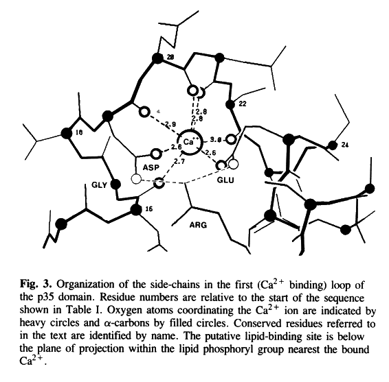
Our work was carried out before structure prediction programmes like AlphaFold took centre stage! I am particularly proud of our prediction of the annexin Ca2+ ion and lipid head group binding site (shown in the images). We were helped by the repetitive nature of the annexin core domains and its presence in all the annexins then sequenced, which reinforced confidence in our structure predictions.The annexin and lipid binding sites were confirmed experimentally by the x-ray crystallographic study (of recombinant rat AnxA5). Much later my own institute set up an x-ray crystallographic facility which I would have been delighted to collaborate with, but I sadly I had moved to an industrial post by that time.

There have been a very large number of immunolocalisation studies of specific annexins in many different organisms: too numerous to reference here. Needless to say we made many studies of our own. I include a few striking images below

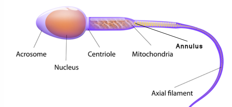
Bovine sperm. This is a highly simplified type of terminally-differentiated cell with an easily distinguished internal structure. Here the main features have been indicated. Sperms are set up for a short trip with fully activated (torpedo-like) functions:- Oocyte sensors are present in the head piece plasma membrane; an acrosomal "cap" containing enzymes which penetrate the egg membrane post fusion (the acrosome is a sort of specialised secretion organelle); a haplotype genome within the nucleus below the acrosome; a power station of stacked mitochondria which supply energy to the end piece and are separated from the motile axoneme made up of parallel microtubules and dynein protein arms by a cytoskeletal barrier known as the annulus. I stained for annexins hoping to see annexin cytolocations more clearly in this cell type. Certainly AnxA2, AnxA4 and AnxA6 detected were clearly associated with different sperm structures. Other workers have examined annexins in sperm, but this work remains to be followed up in terms of functions. The images below each show the same sperm in phase contrast and immunofluorescence.


Since their discovery, the literature on annexins has grown extensively. Many questions have arisen about both their biology and biochemistry. One striking example is that all annexins lack signal sequences that would allow them to be secreted by the conventional ER/Golgi exocytotic pathway; yet they appear in and have roles in blood plasma. The mechanism of release appears to be real and not just due to damaged cell cytosol leaking. It has been termed unconventional or anomalous secretion and this is rare but not unique to the annexins.
Annexin genes have been identified by genomic sequencing in all eukaryotic organisms so far except yeast, but are apparently absent from prokaryotes. There are 12 human annexin genes ranging in size from 15kb to 96 kb. Their tissue distribution has been most intensively studied in mammals. Extensive studies using gene knock out (KO) or knock down (KD) have been carried out in mice to try to assess function (Grewal et al. 2021).
All cells may well express every annexin at some stage of their differentiation or life cycle, but preferential levels have been found in many tissues. This is indicated in the table attached as a guide (but do note that not all published work will be found to confirm the expression levels shown!) Caution is required in interpretation of this since the tissues listed contain a wide variety of differentiated cell types. A number of reviews report that AnxA1 appears widely expressed in somatic tissues and particularly prominent in monocytes: macrophages & neutrophils, the neuronal and endocrine systems. AnxA2, AnxA3 & AnxA4 are prominent in lung, pancreas, colon, ileum and adrenal gland. A5 appears universal in tissue types apart from neurones. A6 is widely expressed in cell types), except in ileal epithelial cells & parathyroid gland. AnxA6 has 8 of the characteristic Type II calcium binding domains. A7 has a uniquely large amino terminal region terminus and is present in alternatively spliced versions: a 47 kD isoform present in all tissues apart from skeletal muscle, and a 51 kD isoform is present in heart, brain and muscle. AnxA8 is expressed in lung, liver, kidney, skin and placenta. AnxA9 was detected first of all in foetal liver and spleen expression libraries. It is highly homologous with AnxA2, but the type II-Ca2+ binding sites in its core domain are not able to bind calcium and its membrane and phospholipid-binding properties are calcium-independent. AnxA10 is mainly found mainly in GI tract epithelia. AnxA11 is widely expressed in tissues with cytoplasmic and nuclear localisation. During the cell cycle AnxA11 appears to translocate to permit midbody formation and the completion of cytokinesis.

Such a wide distribution in eukaryotic cells and tissues suggests that annexins supply critical functions to both multicellular and unicellular organisms. A consensus is gradually emerging that they are proteins which form part of the defence and repair mechanisms essential in all multicellular and many unicellular organisms. In both plant and animal kingdoms many biotic and abiotic stress situations appear to upregulate, translocate and activate annexins. So much so they might be considered an important component of higher organism's innate immune system: some annexins appear to cross the plasma membrane as do cytokines and AnxA1 (for example) binds the formyl peptide receptor to trigger a defensive response toward bacteria. Biochemically they appear to be acting as "adaptor or scaffold" molecules, reversibly bridging between plasma and organelle membranes and cytoskeletal proteins like filamentous actin.
There are reports of specific expression of annexins in the early embryo and annexins may be playing a key role in pattern formation during embryogenesis. The identification of specific proteins which appear to be acting both extracellularly and in the cell cytosol is unusual. My best guess is that, first arising in a very ancient progenitor, evolution has since adapted them for many intracellular and even extracellular tasks. Regulated, reversible membrane lipid binding appears to be an extremely useful property, with so many other membrane-associated proteins being permanently membrane embedded. AnxA13, on genomic considerations, appears to be the closest representative to a very early ancestral molecule. Perhaps the appearance of an ancestral annexin occurred during the transition of nucleated cells to multicellularity or even, like present day slime moulds, promoting reversible aggregation of isolated cells?
Interactions of annexins with cell cytoskeletal proteins have been reported. This is most clear from both in vitro and in vivo observations for AnxA2 which binds F-actin. AnxA1 apparently binds both F-actin and profilin. AnxA5, AnxA6 and AnxA11 also may interact with cytoskeleton-associated proteins. My own cytochemistry of AnxA2 in sperm suggests an interaction with septins or septin associated proteins in the sperm annulus. These interactions with cell cytoskeleton indicates a role as membrane scaffolding. Annexin A5 clearly self-associates to form a 2-dimensional lattice through trimer formation (an analogous property with proteins like clathrin and COPI and COPII proteins that function in intracellular membrane traffic ) Both AnxA1 and AnxA2 have been reported to play important roles in cell division. So Fankhaenel et al. 2023 consider that AnxA1 is a polarity cue orienting the mitotic spindle apparatus for orderly epithelial organisation in mammalian epithelial morphogenesis. This suggests linkage between microtubular elements and plasma membrane. AnxA2 is reported (Benaud et al. 2015) to be important in the initial stage of cytokinesis, presumably by interaction with actomyosin assemblies. Interestingly, Creutz 2023 reports that recombinant AnxA4 rescues cytokinesis blocked in E.Coli by beta-lactam antibiotics.
Annexins are components of the complex and highly regulated molecular "machinery" enabling the directed subcellular transport of membrane in the form of vesicles and their cargo (Gerke et al. 2005). The properties enabling this function include membrane lipid attachment, multiple protein partner recognition and regulated control by kinase / phosphatase action. Key transport pathways include exocytosis and endocytosis at the level of the plasma membrane, but there are many other intracellular membrane transport processes at the level of endosomes, lysosomes and the Golgi / endoplasmic reticulum. Their roles include association with / modulation of specific lipid "raft" composition (Lafont et al. 1998), induction of membrane curvature, supporting membrane - membrane contact through "tethering" interactions and membrane linkage to cytoskeletal elements either through direct binding or through intermediaries like the cytoskeletal accessory proteins as described in the last paragraph.
Tomas Grewal and colleagues provided a recent review of the role of annexins in membrane trafficking.
The figures below give examples of a specific triggered exocytosis followed by the uptake of secretory vesicle membrane. The chromaffin cells illustrated contain relatively large quantities of annexins, especially AnxA1, AnxA2 and AnxA6 which are implicated in these two key processes.
Annexins do seem to be involved in both triggered and constitutive membrane vesicle trafficking, participating in different ways in exocytosis, phagocytosis, receptor mediated and basal endocytosis, vesicle export from the Golgi apparatus and from late endosomes. One of our early EM images of adrenal chromaffin cells stimulated by the adrenergic agonist carbamylcholine, showed secretion granules translocated into the cortical submembrane zone (left image) and, in what we now believe to be the case, using annexins as tethers to the plasma membrane while the exocytotic "machinery" is assembled. Following exocytosis, former granule membrane appears to be recovered in coated pits (right image). AnxA1 may mediate endocytic vesicle fusion and transit through promoting SNARE protein assembly (Song et al. 2023)

Roles for annexins in mitosis - especially AnxA1 and AnxA2 - have been proposed recently. The molecular mechanisms involved remain to be clarified. In tissue formation and repair, oriented cell division is essential for the establishing and maintenance of correct tissue topography - as in epithelial acini for example. AnxA1 has been reported to be necessary for correct orientation of the mitotic spindle in polarised epithelia (Fankhaenel et al.). Knockdown of AnxA1 resulted in disorganised epithelial acini arising from loss of normal (planar) cell division. AnxA2 appears to participate in the early phase of cytokinesis and to be required for correct formation of the cortical cleavage furrow (Benaud et al.). Participation in biomolecular condensates (BMC) BMC represent micron-sized specific associations of proteins and nucleic acids which, unlike many organelles, are not membrane-bounded. These biomolecular collections act as phase-separated liquids. Physical structures include colloidal emulsions, gels, liquid crystals and other aggregates. Examples include nucleoli, Cajal bodies, RNA and stress granules. Grindheim et al. report the presence of membrane-free spherical bodies, co-aligned with astral microtubules, in mitotic cells which form between prometaphase (breakdown of the nuclear membrane) and telophase (reformation of the nuclei of daughter cells). The BMC observed contain endomembranes, but no other recognisable organelles, implicating them as mitotic reservoirs for specific endomembranes or membrane recycling machinery. It is suggested that these peripheral condensates, associated with the twin sets of mitotic astral microtubules, serve the still unexplained underlying origins and inheritance of organelles which must occur in the separated daughter cells. The actin/lipid and mRNA binding AnxA2 is present in these BMC as well as nuclear lamin B. Its role remains to be determined. The importance of BMC and their content of AnxA2 and lamin B in mitotic partitioning is further explored by Grindheim et al.(2023) . These authors find that BMC contain compartments operating in biosynthetic or endocytic membrane recycling (defined by Rab1, Rab11 or endocytosed transferrin) – but lack other membrane organelles. They infer that BMC constitute a mitotic reservoir for selected endomembranes Association of annexins with BMC appears not to be restricted to AnxA1 and AnxA2. Phase-separated membrane-less RNA granules have been shown to "hitch-hike" on endosomes (in particular lysosomes) using AnxA11 as a molecular tether (Liao et al.). The potential association of annexins with other BMC appears highly likely. With their specificity for actin, membrane lipids and nucleic acids, annexins may be expected to participate in many stages of the complex molecular machinery underlying mitosis and the cell cycle. Subject to regulation by Ca2+ ions, phosphorylation and other PMC, the mechanisms of action of annexins in mitosis can be expected to occupy researchers for a long time to come!
Plasma membrane integrity is vital for tissue homeostasis and the function of cells in the circulation. Membrane damage regularly occurs through physical processes (exercise) and chemical/biochemical injuries which include the effects of toxins and infection. The repair process generally requires PS or PIP2) lipids, membrane vesicles and calcium ions. In intact tissues and circulating plasma cells, stresses and injuries are autonomously repaired. AnxA1, AnxA2, AnxA4, AnxA5, AnxA6 and AnxA11/AnxA11a have all been implicated in this process, but other cellular components also form part of the repair machinery. AnxA2 is a component of the Ca2+- dependent repair mechanism in endothelial and muscle cell plasma membrane lesions. This may be a universal response in eukaryotes: analogous healing of laser-induced membrane lesions by AnxC1 - an ortholog of mammalian AnxA7 - in the fungus: Dictyostelium discoideum has been reported. A variety of model or natural isolated cell membranes have been described in which AnxA2 has featured as an annexin recruited at the first, or early stage of cell response to membrane lesions (Kayelo et al. 2023). AnxA6 has been shown to form a Ca2+ dependent — "cap" incorporating membrane and F-actin at sites of plasma membrane disruption mediating resealing, especially in damaged skeletal muscle sarcolemma (Demonbreun et al 2019) and Demonbreun et al 2016 ). This crucial repair process still requires additional research to define it at the mechanistic level. AnxA4 and AnxA5 form trimers at membrane surfaces, inducing high membrane curvature. AnxA3 (and possibly other annexins) do not show this property, which may be related to annexin paralog membrane binding affinity as well as the requirement for heterologous annexin binding sites (Zanjani et al. 2023).
The septin cytoskeleton together with F-actin appears actively involved in this process (Prilusky et al. 2023). It is now frequently reported that annexins such as AnxA2 are important in the repair of tumour cell membranes. Metastatic tumour cells are subject to membrane lesions during circulation. Knockout of AnXA2 has been reported to inhibit such metastasis (see Annexins in Medicine section of this web). Secretion of exosomes appears coupled to Ca2+-dependent plasma membrane repair (Williams et a. 2023). In both intact and permeabilised cells, AnXA6 appears to be recruited to Multivesicular Bodies (MVBs) and is required for Ca2+-dependent exosome secretion. ANXA6 depletion leads to peripheral accumulation of MVBs. ANXA6 truncations localize to internal membranes, suggesting that ANXA6 may tether MVBs to the plasma membrane. Cells secrete exosomes and other extracellular vesicle (EV) types upon plasma membrane damage. Such plasma membrane repair-induced EV secretion may contribute to the pool of EVs present within biological fluids.
The Endosomal Sorting Complexes Required for Transport (ESCRT) machinery is composed of 4 cytosolic proteins. It is required for processes including multivesicular body formation, cell abscission and viral budding. Also involved in membrane repair such as in damaged lysosomes. AnxA7 is required for ESCRTIII -mediated plasma membrane repair (Sønder et al. 2019 ), but AnxA1 and AnxA2 can act independently of the ESCRT complex for repair of damaged lysosomes ( Yim et al. 2022 )
Heffner et al. (2024) showed differences and interdependencies in the recruitment of ESCRT-III and Annexin proteins to sites of membrane damage. Annexins are recruited immediately, ESCRT-III assembles only after membrane sealing. Mammalian cells express 11 different ESCRT-III subunits called Charged Multivesicular Body Proteins (CHMPs). ESCRT-III sheds damaged cell membranes and Frontotemporal Dementia (FTD) and Amyotrophic Lateral Sclerosis (ALS) associated mutations in CHMP2B and ANXA11 compromise the repair process. These data present an integrated ‘sealing and healing’ model of events allowing membrane repair and restoration of membrane integrity.
Another study reported a previously unrecognized function of AnxA7 in lysosome repair. This repair process operates independently of the Endosomal Sorting Complex Required for Transport (ESCRT) machinery. AnxA7 plays a role in repairing damaged lysosomes, different from its role in repairing the plasma membrane, where it facilitates repair through the recruitment of ESCRT-III protein components (Ebstrup et al. 2023 ).
Extracellular AnxA5 has been shown to participate in the formation of multinuclear osteoclasts. As such it appears to be involved in the cell fusion stage of osteoclastogenesis. Chernomordik and Melikov examined cell fusion leading to multinucleated osteoclasts and concluded that AnxA5 and S100A4 complexes supported by the glycoprotein syncytin 1 (Syn-1), assembled at externalised PS drove close approach and fusion. It was suggested that the first annexin identified: AnxA6 was involved in biological membrane fusion processes. We now know that membrane fusion is supported by annexins, but other proteins are essential for the bilayer fusion stage.

Post-transcriptional regulation of gene expression is markedly affected by non-coding RNAs especially microRNAs (miRNAs) of which more than 1000 have been identified. The expression of miRNAs 196a, 26b and 562 is inversely correlated with the level of cellular AnxA1 and miRNA324 by AnxA2. The annexins appear to regulate the synthesis, localisation and transport of the regulatory miRNAs. Considering the intimate involvement of annexins in tumour cell growth, proliferation and metastasis, these feedback loops are highly significant.
Annexins have been recognised as RNA binders. Tartaglia et al. (2025) used a computational approach to predict RNA sequences that participate in this interaction. Some annexins: especially AnxA7 and AnxA11 exhibit strong phase separation properties. The authors suggest that these phenomena play a role in RNA trafficking and localisation - especially in the axonal bodies of neurons.
AnxA11 participates in a (reversible) liquid-liquid phase transition which requires the intrinsically disordered proline-rich N-terminus (33% pro). This region of AnxA11 is required for the incorporation of RNA into granules and leads to the hitch-hiking of RNA granules complexed with lysosomes along microtubules. AnxA11 is acting as a temporary tether in this transport process. Dudas et al. (2024)

Annexins have been “fingered” for involvement in so many different intracellular and extracellular activities (the list is probably still rather incomplete) that design of annexin-directed therapeutics to target specific pathologies presents considerable challenge. In a number of diseases, dysfunctional expression and/or translocation of specific annexins is highly correlated with poor outcomes, meaning that the proteins are considered potential biomarkers. Unresolved inflammation is a major contributing factor to tissue damage and mortality. AnxA1 is widely reported as a systemic responder to the resolution of inflammatory conditions. Peptide mimetics (such as the AnxA1 amino terminal peptide: Ac2-26) have been widely used in animal models of inflammation, but this field still remains quite open. The blanket term "annexinopathies" was coined following detection in the late 1990s of aberrantly raised levels of AnxA2 in acute promyelocytic leukaemia (Menell et al. 1999 ) as well as other conditions linked to abnormal annexin expression. Also see Rand, J. 2000. The long established involvement of AnxA1 and AnxA2 in the inflammatory response, fibrinolysis and more recently in cell membrane repair, has led researchers to look for their expression in many normal and pathological conditions. Annexins are frequently reported to be biomarkers of various pathologies. Of current focus, annexins and peptide mimetics are under consideration as possible therapies. With Brazilian co-authors, I have very recently reviewed the molecular cell biology of the annexins most frequently cited in relation to human pathologies (de Souza Ferreira et al. 2023). As can be seen below, AnxA5 in particular would appear to be implicated as a therapeutic in many inflammatory and other situations where cell surface exposure of the normally internal phospholipid phosphatidyl serine is exposed on epithelial or endothelial cell surfaces.
This section of the webpage will doubtless need to be updated frequently!
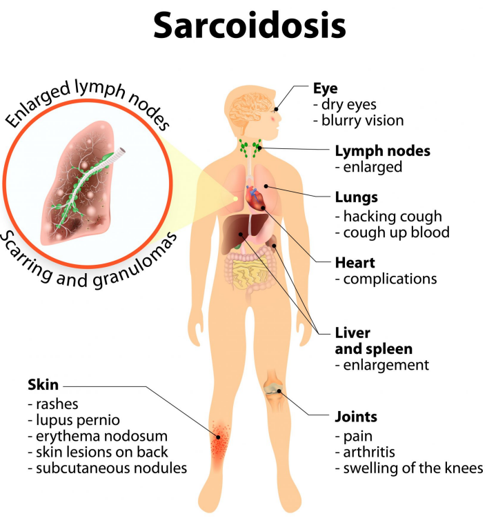
Diagnostics | Brain | Vision | Pregnancy | Infection | Diabetes | Autoimmune | Pneumonia | Atherosclerosis | Acute liver failure | Sarcoidosis | Cancer & tumorigenesis | Inflammatory Bowel disease | Osteoporosis | SARS-Cov-2 & sepsis | Alzheimer's | Cardiovascular disease & stroke
One of the earliest applications of the annexins was for detection of the exposure of the phospholipid phosphatidylserine (PS) in the external leaflet of cell membranes. In normal cell metabolism PS is asymmetrically resident in the cytoplasmic leaflet, retained there by an ATP-dependent flippase. In platelets it can be exposed on the extracellular surface by cell-type dependent scramblase action ( Nagata et al. 2016). More generally, extracellular PS exposure is taken as an indicator of plasma membrane damage or cell death (apoptosis). It has come to be regarded as an "eat me" signal to phagocytes for tidy clearance from the circulation as well as creating a scaffold for clotting factors on activated platelets. Erythrocyte aging also leads to surface exposure of PS which constitutes an active trigger for cell removal from the circulation.
AnxA5 has been derivatised with fluorescent markers (Fluorescein isothiocyanate) or short lived radioisotopes and has become an almost universal detection agent for apoptosis in fundamental research and clinical medicine following chemotherapeutic treatment (Schaper and Reutelingsperger).

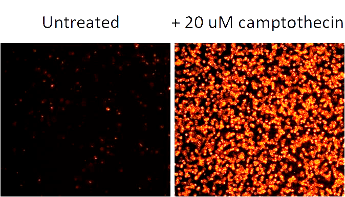
mRNA in axons via ribonucleoprotein complexes (RNPs) is necessary for neuron function and survival. Feng et al. (2025) identify Annexin A7 (ANXA7) in the transport of T-cell intracellular antigen 1 (TIA1)-containing RNPs by linking them dynein. Persistent axonal Ca²⁺ elevation disrupts ANXA7’s linker role, causing the detachment of TIA1 granules from dynein, consequently impairing transport and triggering pathological TIA1 aggregation within axons. Similarly, ANXA7 knockdown decouples TIA1 granules from dynein, severely obstructing trafficking and causing pathological aggregation of TIA1 in axons, which culminates in axonopathy and neurodegeneration both in vitro and in vivo. Conversely, ANXA7 overexpression enhances trafficking and counteracts aberrant aggregation of TIA1-containing RNPs in axons. Our findings elucidate a novel mechanism underlying RNP axonal transport, highlighting its significance in the biology and pathology of central neurons.
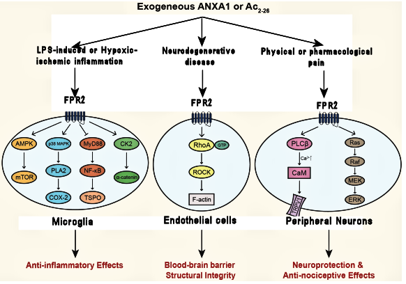
AnxA1 is important for maintenance of homeostasis especially anti-inflammation in neurological tissues (brain and eye) It is observed to maintain neurological homeostasis through G protein linked formyl methionyl receptor (FPR2) signalling but probably also via other routes.Its neuroregenerative potential makes it interesting in development of neuroprotective / neuroregenerative approaches. At the level of the Blood Brain Barrier (BBB), AnxA1 has been detected in the microvascular endothelial cells as well as the associated mural support cells (pericytes and smooth muscle cells), the microglia and the ependymal cells lining the spinal chord and CNS ventricles. AnxA1 makes a variable appearance on neurons themselves. Nuclear translocation of AnxA1 (in S100A11-free form) in CNS cells via the nuclear transport protein importinß has been shown to trigger apoptosis via a p53/Bid/caspase-3/polyADP-ribose polymerase pathway (Li et al. 2016). Oxygen/glucose deprivation of neuronal cells leads to phosphorylation of AnxA11 resulting its translocation to the nucleus and triggered expression of the BID gene, where caspase-mediated neuronal apoptosis can follow. For a recent review: (Wang et al. 2023))
Philipe de Souza Ferreira et al (2025) have reviewed the action of AnxA1 in neurological disorders, focussing in neuroprotection and glial cell modulation. The authors suggest that a comprehensive understanding of the intricate mechanisms of ANXA1 in neurons and glial cells is leading toward therapeutic strategies to alleviate neuronal damage in neurological diseases.
Back to menu . .
The pathogenesis of serious eye diseases involves the vasculature. Conditions include retinal vein occlusion (RVO), diabetic retinopathy (DR) and wet age-related macular degeneration (AMD). Presently the only effective therapy available (for RVO) is intra injection of antibodies to Vascular endothelial growth factor (VEGF) to reduce occlusive overgrowth. Aberrant signalling by phosphatidylserine (PS) present in the outer lipid leaflet of endothelial cells provides an alternative target for defective vascular therapies. Annexin A5 binds with high affinity to PS, is normally present in the extracellular space and lacks many of the other properties and functions of other annexins which could drive unwanted side effects. Recombinant AnxA5 is presently in clinical trials for RVO and there are many other indications for specific therapies related to exposure of cell surface PS. This topic is further explored in the section on Pharmaceuticals development below
In the distressingly common age-related macular degeneration (AMD), serum AnxA3 levels were found to be significantly higher in wet AMD than in control groups (Ozturk A et al. 2023). These authors suggested AnxA3 could be used as a diagnostic and potentially therapeutic approach to AMD.
The pathophysiology of annexins in retinal vascular diseases - particularly AnxA5 - is explored in detail by Anna Frostegård and Anders Haegerstrand in a recent review in Pharmaceuticals (July 1994)

Annexins are involved in diverse cell functions including: response to pro-inflammatory conditions; blood coagulation and complement reactions; apoptosis, membrane trafficking in endo and exocytosis; intracellular communication; extracellular matrix and cytoskeletal rearrangements, signal transduction (both as inducers in the extracellular space and effectors within the cytoplasm itself) and membrane resealing. From their involvement in such processes they are beginning to be thought of as components of our innate immune response. It would certainly be expected that they are strongly implicated at various stages in the complex cell processes which underlie pregnancy, especially in the critical stages of blastocyst (early embryo) implantation and the subsequent morphological changes of the surrounding uterine wall (see image above). Controversially in vivo studies using annexin gene knock-down (KD) and knock-out (KO) mouse models suggest that removal of individual annexins AnxA1, AnxA2, AnxA4, AnxA6 or AnxA7 did not appear to impair the key attributes of normal animal cell biology, suggesting redundancy within this protein family. However, annexin knockout mice were phenotypically distinct under conditions of stress. But there does remain some controversy about the results of annexin KO in the literature and this is addressed in the reviews mentioned in this section.
Loss of AnxA5 had been considered to lead to pregnancy failures associated with antiphospholipid syndrome and thrombosis (see Rand et al., 2010 and later in this section). Studies have found that anti-AnxA5 autoantibodies are a risk factor for reproductive failures (recurrent abortion) See Murad et al. 2023. Reduced litter size and increased foetal loss in AnxA5 KO mice has been reported (Ueki et al. 2012) and smaller foetus size was apparent upon maternal loss of AnxA5. This could be reduced by administration of the anti-coagulant heparin to prevent clotting in the placental circulation. These authors suggested that AnxA5 KO animals might be an appropriate model for investigations of thrombus formation causing infertility by the antiphospholipid syndrome in humans. Annexins have been more extensively explored by Grewal T. et al. (2016 & 2021). These found that conception, embryogenesis, cell differentiation, tissue development and function, and fertility were not substantially affected (even in some cases, where two annexins were removed). It remains to this day as an hypothesis that functional complementation by unaffected annexins or other, presently unidentified factors, may provide answers to this apparent conundrum.
However, annexins appear to show themselves in pathological circumstances of pregnancy (as indeed they do in many other situations of stress where they have been studied in the animal kingdom!) High levels of AnxA1 are expressed in the human uterus. The underlying rationale for actions of AnxA1 in utero, the crucial processes of blastocyst implantation and subsequent changes to the endometrial cellular environment likely relates to the well established anti-inflammatory properties of this annexin. Its transmembrane signalling via the FPR2 receptors has multifunctional and cell-type specific functions, such as cytoskeletal rearrangement via the recruitment of intracellular annexins including AnxA1 and AnxA2.
AnxA1 levels are correlated with pathologies of pregnancy especially gestational diabetes mellitus and pre-eclampsia (PE). This has been directly demonstrated in mouse models: Anx A1 -/- mice were found to have increased uterine inflammation at the stage of implantation and a reduction of successful pregnancies. Increased levels of AnxA1 and ultrasensitive C-reactive protein were found in plasma of women diagnosed with PE (Perucci et al. 2015). These also showed elevated levels of autoantibodies against AnxA1 and abnormal levels of inflammation.
Other annexins also play an important role in early pregnancy. A maternal deficiency of AnxA2 expression was found to influence aberrant decidualization and shallow cytotrophoblast invasion, suggesting that decidualization resistance could underlie shallow trophoblast invasion and the poor establishment of the maternal-foetal interface. (Ruikar et al. 2021) Shallow cytotrophoblast invasion through the uterine decidua into the spiral arteries is implicated in the pathogenesis of PE, although the cause of the deficient arterial invasion remains unknown.
AnxA4< has been considered to promote trophoblast invasion via the PI3/Akt/eNOS signalling pathway (Xu et al. 2019). Overexpression promoted cell proliferation and invasion while knockdown (in rodent models) had the opposite action.
On the plasma surface of the placental syncytiotrophoblast it has been established that expression and translocation of AnxA5 to the apical (maternal blood facing) surface sets in place a dynamic "shield" against the coagulatory cascade that otherwise accompanies the recognition by clotting factors of cell surface exposed phosphatidyl serine (PS).
The antiphospholipid syndrome (PE) is characterized by antibodies directed against phospholipid-binding proteins and phospholipids attached to cell membrane receptors, mitochondria, oxidized lipoproteins, and activated complement components. When antibodies bind to these complex antigens, cells are activated and the coagulation and complement cascades are triggered, culminating in thrombotic events which negatively affect foetal nutrition leading to the pregnancy morbidity that further define PE syndrome (reviewed by Green D. 2022). The phospholipid-binding proteins most often involved are AnxA2 and AnxA5, β2-glycoprotein I, prothrombin, and cardiolipin A.
A present "hot topic" of both fundamental and translational research is the shedding of extracellular vesicles (EVs) either as exosomes formed through secretion of multivesicular bodies (MVBs) or ectosomes which arise as blebs formed from cell plasma membrane. These are presently being regarded as an important component of intercellular communication, their cargo of key relevance for biomarkers of disease and as potential carriers for exogenous therapeutics. It's possible that annexins are involved in the formation and transport of EVs and may represent cargo associated with them. Biomolecules and EVs shed from placenta (the placental secretome) in normal and pathological situations (such as PE) has been reviewed by Aplin et al. (2020). These authors speculate that cross-linking of proteins in the secretome may expedite their removal by blood phagocytes.
Pathogens (microorganisms and viruses) recognise host tissues through cell-surface associated molecules which may range from glyco-conjugates to membrane proteins. Infection generally follows either a stable adhesion process - the pathogen remains anchored in the extracellular space - or receptor recognition followed by entry by an endocytic route. Once inside cells, pathogens may manipulate vesicles or membranous tubules in recycling endocytic pathways, avoiding endolysosomes, to transfer genome into the cytosol or take up residence within intracellular membranes engineered to avoid the subcellular immune response.
The role of annexins both within parasites and their hosts during infections has been reviewed in depth by Rashidi et al. (2023). These authors indicate that parasites (especially helminths) express and secrete annexins in pathogenesis and modulation of host annexins could be employed as a survival strategy by intracellular protozoan parasites. Expression of AnxA2 appears ubiquitous across multiple cell types. The impact of AnxA2 on viral life cycles is reviewed by Park et al. (2024). These authors note that it remains unclear how AnxA2 and the virus associate to regulate virus life cycles at the molecular level.

Diabetes, obesity and connected disorders can be collectively considered under the heading of metabolic dysfunction which can arise through disturbed cell homeostasis. As an antigen expressed on cell membranes, AnxA2 initiates local inflammation and damage through binding to auto-antibodies. Aberrant expression of AnxA2 has been found in numerous kidney diseases. AnxA2 has been shown to act as a co-receptor of integrin CD11b mediating NF-kB-dependent kidney inflammation, which is further amplified through AnxA2/NF-kB-triggered macrophage M2 to M1 phenotypic change. It also modulates podocyte cytoskeleton rearrangement through Cdc42 and Rac1/2/3 Rho pathway causing proteinuria. Thus, AnxA2 is implicated in the pathogenesis and progression of various kidney diseases. The expression and roles of AnxA1, AnxA2 & AnxA6 in glucose metabolism and adipocyte function have been reviewed by Grewal et al. Inflammation and altered metabolic activity underlie diabetic nephropathy. AnxA1 appears to regulate lipid metabolism in proximal tubular epithelial cells. Once again suggesting that it may be a useful therapeutic target for this condition.
AnxA1 is considered to have significance in patients with type 2 diabetes mellitus with foot ulcers Overall, the lower levels of anti-inflammatory AnxA1 in diabetic foot ulcers indicating its role in the inflammation homeostasis required for the initiation of wound healing (Shwetha et al. 2023)
AnxA5 associates with PS-rich extracellular leaflet of the trophoblast plasma membrane: most cell membranes do not display PS in their outer cell phospholipids in surface membranes. The reason appears to be to raise a protective barrier to inhibit coagulation that would otherwise occur at exposed PS or other negatively charged phospholipid sites. Plasma antiphospholipid antibodies break down the protective barrier, displacing AnxA5, accelerating coagulation of plasma exposed to to trophoblasts and endothelial cells with potential pregnancy loss (Rand et al.).
Endometrial receptivityAnxA7 appears to have a role in the regulation of endometrial receptivity and embryo implantation. In women suffering repeated pregnancy loss, AnxA7 RNA transcripts and protein levels are decreased in the midluteal window of implantation (Alauddin et al.)
InflammationAnxA1 was one of the first members of this family to be investigated for its resolving action on the inflammatory response. Acting at least through interaction with the formyl peptide receptor (FPR2) on endothelia, its actions are pleiotropic. An important site of interaction is represented by the amino terminus and peptides comprised of just 23 or so amino acids mimics many of AnxA1 actions. This has led to the exploration of mimetic peptides in anti-inflammatory therapies. The anti-inflammatory actions of AnxA1 has received many literature reviews: one recent example: its action in Inflammatory bowel diseases - is given here (Xu et al. ))
Periodontitis is an inflammatory condition that affects the tooth‐supporting structures (incl. periodontal ligament cells (PDLCs). Damage is arises from the host's immune response toward bacterial deposits around the teeth. Luo et al. (2024) considered that AnxA1 has a protective effect against lipopolysaccharide (LPS)‐induced inflammatory responses and cellular damage . AnxA1 demonstrated potent anti‐inflammatory effects by inhibiting the secretion of interleukin 6 (IL‐6), interleukin 8 (IL‐8), and monocyte chemoattractant protein‐1 (MCP‐1). The authors found that AnxA1 protected PDLCs against LPS‐triggered inflammation and cell senescence by activating sirtuin (SIRT1) signal pathway. Their findings indicated that AnxA1 could serve as a promising therapeutic strategy for the treatment of periodontitis.
Back to menu . .Regeneration of liver tissue is often overwhelmed leading to acute liver failure. A new AnxA2+ migratory hepatocyte lineage was noted by Matchett et al. (2024) Knockdown of hepatocyte AnxA2 reduces HGF-induced human and mouse hepatocyte migration in vitro, preventing necrotic wound closure following induced mouse liver injury. The study revealed unanticipated aspects of human liver regeneration, identifying a novel migratory hepatocyte subpopulation mediating wound closure following liver injury. The authors suggested that interventions designed to promote hepatocyte migration with rapid reconstitution of normal hepatic microarchitecture and repair of the gut–liver barrier may open up a new area of therapeutic discovery in regenerative medicine.
Back to menu . .Annexins are becoming more frequently used as biomarkers to detect or determine prognosis of disease. In community aquired pneumonia (CAP) Gu et al. 2023 found that plasma AnxA1 levels correlated with a significantly higher survival rate in patients with severe CAP.
Back to menu . .
In recent years, annexins have become strongly implicated in many aspects of cancer and the literature has expanded substantially. Very generally speaking, abnormal under- or over-expression of various annexins correlate with different aspects of carcinogenesis. This has been most widely studied in breast cancer, but many other tumours have also been investigated. A recent comprehensive analysis relating to breast cancer: (Fathi et al. 2023) exemplifies this. Also a "pan-cancer evidence of prognosis, immune infiltration, and immunotherapy efficacy for annexin family using multi-omics data" has been published ( Shen et al. 2023).
High levels of AnxA1 expression are associated with poor prognosis of a number of cancers. (Al-Ali et al. 2024) find that a humanised monoclonal targeting AnxA1 exerts an antiproliferative effect. It disrupts AnxA1 binding to FPR1/2 receptors and leads to arrest of metastatic cells at the G1 phase of their cell cycle. Better known for its anti-inflammatory action, AnxA1 is also a mediator in several signalling pathways (eg PI3K, MAPK/ERK and STAT3) which are implicated in tumour progression. It is suggested that antibody targeting of AnxA1 in patients with tumours overexpressing AnxA1 may offer a therapeutic intervention.
The interaction between immune cells and surface-exposed phosphatidylserine (PS) on the surface of apoptotic cells,from chemotherapies, contributes to the formation of an immunosuppressive tumour microenvironment (TME) - hindering tumour cell interaction with immune cells. AnxA5 administration rescues the TME immunosuppressive state. Conjugation of a tumour-antigen peptide with AnxA5 upregulates the peptide's immunogenicity and antitumor efficacy when administered after chemotherapy. This therapeutic antitumor effect of an AnxA5-peptide fusion can be further enhanced by administration of other immune checkpoint inhibitors. Our findings support the administration of AnxA5 following chemotherapy is a promising immune checkpoint inhibitor for cancer treatment. (Kang et al. 2020).
AnxA2 has been a particular focus in tumorigenesis and consequences of its under or over expression have received wide attention in the literature. In consequence, diverse therapies (both small and macromolecular drug entities) have been suggested or tested. One recent example is the naturally occurring flavonoid Baicalin which was shown to target AnxA2 with anti-tumour and apoptosis-inducing actions.
In light of its immunomodulatory and ant-inflammatory actions, AnxA1 has had even more studies in relation to cancer than AnxA2. Both Annexins play a prominent role in plasma membrane repair, which is a function important in circulating metastatic cells which are subject to physical damage in the circulation. This is critically reviewed "AnxA1: A blessing or a curse?" ( Foo et al. ). AnxA7 and its co-related tumour cell death silencer protein (suppressor of death domains SODD) appear to regulate a migratory phenotype of liver cancer cells. Down-regulating AnxA7 in Cha-P cells decreased proliferation, migration and invasion of tumour cells, and SODD expression was decreased (Wang et al. 2023) Proteomic analysis has identified AnxA1 as a potential biomarker of thyroid cancer malignancy, with its levels increased in malignant samples. Also upregulated were the acetylated peptides of AnxA1, revealed by the peptidome analysis (Coelho et al. 2023)
AnxA7 (the first annexin identified, then known as synexin) potently aggregates lipid bilayers in a calcium and GTP dependent manner. AnxA7 is associated with tumour gene suppressor properties. Srivastava et al. 2023 found a dominant-negative AnxA7 triple mutant led to the loss of lipid membrane binding activities and impaired signalling, enhancing the proliferation of prostate cancer cells
This area is almost continuously reviewed! For a recent survey see Zhang et al. 2023

Exposure of phosphatidyl serine on the surface of colon vascular capillary is a circumstance leading to inflammation of this component of the vasculature. It has been reported that AnxA5 targets the colon and relieves experimental colitis (Zhang et al. 2021)
Back to menu . .Su et al. (2023) Report that AnxA5, which appears to be an important component of the membraneous bone matrix vesicle, plays a key role in bone matrix homeostasis in the deterioration of osteoporosis. It was sharrply deceased in bone matrix in a mouse osteoporosis model.
Back to menu . .AnxA1 appears to be a key modulator in lung inflammation seen in SARS-Cov-2 infection. Resende et al. (2025) Using a mouse model show that endogenous AnxA1 exerts a protective effect. The Mimetic peptide Ac 2-26 (which binds to the AnxA1 receptor) exhibited similar levels of protection. Sepsis, in spite of lengthy and ongoing international research, remains a very seriously challenging condition. Severe SARS-Cov-2 is analogously challenging for urgent treatment in intensive care situations. The established pro-resolving anti-inflammatory, anti-apoptotic, and anticoagulant activities of AnxA1, AnxA2, and AnxA5 appear to offer potential as therapeutics in both circumstances Review: (Mui et al. 2021). This potential is presently being explored by at least two pharmaceutical companies (see later).
Back to menu . .Bartolome et al. (2020) have found that AnxA5 prevents amyloid-β-induced toxicity in choroid plexus, where deposits are formed. Experimental addition of this annexin to choroid plexus cell cultures restored the Aβ-induced impairments on autophagy flux and apoptosis in a calcium-dependent manner
Back to menu . .de Jong et al. (2018) using mouse models, found that AnxA5 reduces infarct size and improves cardiac function after myocardial ischemia-reperfusion injury by suppression of the cardiac inflammatory response
Ischemic stroke is a leading cause of disability and death worldwide. Hu et al. (2025) report that plasma level of Annexin A5, not Annexin A1 or A2, was upregulated in stroke patients when compared to controls. In normal mice, the highest level of Annexin A5 were detected in lung tissues compared with other major organs and lowest level in brain. The authors suggest Annexin A5 may alleviate infarct area and improve general neurological performance following cerebral ischemia. Increased Annexin A5 may derive from lung tissue and permeate across the blood brain barrier to provide a neuroprotective function. Hence Annexin A5 may potentially serve as a therapeutic candidate for defending against ischemic stroke-induced brain injury.
 Currently the Swedish biotechnology company ANNEXIN PHARMA has recombinant AnxA5 in clinical trials for Retinal Vascular Occlusion (RVO).Blood clots form at sites of inflamed and/or damaged endothelial cells which provide the lining of all our blood vessels. In the eye, Central retinal vein occlusion (CRVO) is the blockage of the main retinal vein and branch retinal vein occlusion (BRVO) represents blockage of one of the smaller branch veins. (see the figure below). If persistent (especially in older people) the swelling due to vascular fluid leakage at the centre of the retina (macula) can cause permanent loss of central vision. Swelling of the retinal artery presses on the retinal veins that all exit in the same area. Presently medicines such as anti- vascular endothelial growth factor (anti-VEGF) or steroids may be are used to combat ocular swelling and leakage. These medicines have to be periodically injected into the eye which entails obvious anxiety, discomfort and work absences. Systemic AnxA5 administration is aimed to suppress these localised blood clots without intraocular injection. Annexin Pharma are also investigating its potential in other vascular diseases (see below).
Recent preclinical studies have shown that AnxA5 inhibits proinflammatory responses and improves organ function and survival in rodent models of sepsis. In assessments of the use of AnxA5 in patients with severe COVID-19 a preclinical trial showed that AnxA5 was cleared successfully from plasma with no alteration to blood coagulation (Tschirhart et al. 2023)
AnxA5 is presently seen as an immune checkpoint inhibitor and tumour-homing molecule for cancer treatment (Kang et al. 2020).Tumour and apoptotic cells (sick and dying) externalise intracellular phosphatidyl serine (PS). Tumour cells especially display PS through poor vascularisation and chemotherapies. PS has widely been used as a marker for this. However, unlike phagocytosis of pathogens, the process of apoptotic cell clearance is immunologically silent, avoiding the normal inflammatory response. But this process (efferocytosis) contributes to a localised immunosuppressive environment which can be exploited by tumour cells to avoid immune attack. Cell surface molecules (usually proteins) which normally act to suppress immune attack are known as "checkpoints" AnxA5 binds membrane externalised PS with high affinity hence can act as a so called "immune checkpoint inhibitor" hence can increase the effectiveness of anti-tumour therapies.
As AnxA5 rapidly combines with the membrane lipid phosphatidyl serine (PS) and probably other acidic phospholipids normally not exposed on healthy endothelial cells, its therapeutic use is indicated on other conditions. PS also appears on aging erythrocytes, which of course lack metabolism. Normally these senescent cells are phagocytosed by macrophages and cleared from the circulation. But in sickle cell disease (SCD) crisis events a large number of erythrocytes are senescent, can adhere to endothelial cells and can overwhelm this clearance system. The high level of exposed PS acts to initiate clot formation. Administration of AnxA5 binds to the exposed PS and administration may be indicated to calm the crisis and minimise coagulation (Kennedy 2015).
Sepsis and COVID-19.AnxA5 has recently been directly proposed as a drug candidate to reduce the damage observed in the vascular system and lungs of patients with severe COVID-19 disease.
Annexin A1 Remoter Pharma (Holter, Denmark) is exploring the application of RTP-026, a drug derived from human AnxA1, for inflammation-driven disease such as myocardial infarction.
The first clinical trial of MDX-124 (Med annex Limited, UK), a humanised monoclonal antibody that targets AnxA1 in patients with advanced cancer, is ongoing to find an effective dose of the study drug for future studies. It is based on the critical role of the overproduction of AnxA1 in how certain cancers behave.
Remoter Pharma (Holter, Denmark) is exploring the application of RTP-026, a drug derived from human AnxA1, for inflammation-driven disease such as myocardial infarction.
The first clinical trial of MDX-124 (Med annex Limited, UK), a humanised monoclonal antibody that targets AnxA1 in patients with advanced cancer, is ongoing to find an effective dose of the study drug for future studies. It is based on the critical role of the overproduction of AnxA1 in how certain cancers behave.
Anti-ANXA1, anti-ANXA2, and anti-ANXA5 autoantibodies appear significant for phenotyping and risk stratification, including in seronegative antiphospholipid syndrome. Overall, annexins and their autoantibodies represent promising biomarkers and therapeutic targets; however, the heterogeneity of assays and the limited availability of prospective multicenter data currently hinder clinical translation. Annexins and autoantibodies have recently been reviewed (2026) by Zhou et al.
Plant genomes sequenced to date code for between 8 and 25 non-redundant annexin genes ( Wu et al. 2022) . The typical plant amino acid sequence homology with animal annexins is generally less than 50% and comparison of different annexin sequences from the genome of one species shows considerable variability, but all retain the carboxy-terminal annexin “core” annexin signature key to lipid membrane binding. That is four 70-amino acid internally homologous chain repeats which form 5-fold alpha helical protein domains which carry the annexin “Type II” calcium-binding loops. In plants these loops generally appear in domains I & IV. The first domain carries a conserved tryptophan residue in the “AB loop” which is exposed in the few experimental 3D structures known and hence is available for interaction with the hydrophobic environment of cell membranes. Functions of plant annexins have been well reviewed by Mortimer et al. 2008 and Konopka-Octupolar et al. 2011
The first 3D structure of a plant kingdom annexin was from a bell pepper (Hofmann et al. 2000) and later, from cotton (Hofmann et al. 2003). These confirmed the presence of the canonical annexin cores structures, but a cotton annexin also possesses a sulphur cluster “S3“ formed from methionine and cysteine residues. This is considered to be a structural correlate of a protective property of the annexin through electron transfer to an oxidising agent. It's thought to contribute to the plant’s response to oxidative stresses arising from conditions like heat shock, salinity, nutrient deprivation, tissue injury etc. In contrast with animal annexins, amino terminal sequences which precede plant annexin structural cores are typically relatively short. Although like animal annexins these are highly variable, their short length would not appear to offer much variety to interact with diverse binding partners. However, as with animal annexins, the amino termini do appear to exert allosteric effects on cell membrane binding.
Plant annexin cores bind selectively to the acidic phosphatidyl lipids: phosphatidyl serine (PS) and phosphatidyl inositol's (PI, PI2) and phosphatidic acid (PA) depending upon annexin type and/or ionic conditions (Ca2+ / pH) and produce aggregation of extracellular vesicles and liposomes in vitro. A broader membrane contact surface differentiates plant from mammalian annexins. As in vertebrate annexins, hydrophobic characteristics of this surface (for example, a conserved exposed tryptophan residue in the first plant annexin domain) appear to contribute to both Ca2+ dependent and independent membrane association. Partial membrane insertion is still speculatively reported in the annexin literature and there remains the established fact of annexin translocation, rather than “conventional” ER/Golgi secretion of both plant and animal annexins to the extracellular space.
Oligomerisation on the plasma membrane is a characteristic of both plant and animal annexins and has been considered a response to membrane injury. Given the propensity of particular annexins for self association on lipid membranes, calcium independent membrane association might lead to co-operative assembly of different annexins on cytoplasmic membrane surfaces. Plant annexins are subject to post-translational modifications like animal annexins. They are substrates for specific serine and threonine cytoplasmic kinases and phosphatases which hence exert regulatory controls. Myristate conjugation and glutathionylation (of cysteines) has also been reported. Plants appear to lack typical protein tyrosine kinases, but proteins phosphorylated on tyrosine residues are found in plants, arising perhaps from dual specificity kinase action. At the time of this perspective no plant annexins appear as substrates for tyrosine phosphorylation but that may change in future. The same observation applies to ubiquitinylation and sumoylation although both these PTMs occur in plant cells.
Although not all annexin genes in every plant species are necessarily expressed, the number of potential proteins, not even allowing for alternatively spliced isoforms, are considerable and suggests there remain many functions still to be identified. As with animal annexins, plant orthologs are synthesised in the cytosol and observed to associate both with plasma and organelle membranes. Annexin expression is strongly related to plant developmental stages. (e.g. leaf tip growth and cell elongation: Hofmann et al. 2000). As with animal annexins, plant annexins are strongly implicated in membrane organisation and vesicular transport (Konopka-Postupolska & Clark 2017). They exhibit Ca2+ channel regulation, peroxidase and ATPase/GTPase activities. Tables of other suggested plant annexin functions are starting to appear in recent reviews (Taneja & Upadhyay 2021 and Wu et al. 2022). The table shown below was adapted from the latter two publications.
| Species | Gene name | Function(s) |
|---|---|---|
| Arabidopsis thaliana (Thale cress) | AtANN1 | Wound response, Cold & Heat tolerance |
| AtANN1,2 | Fungal defense, primary root growth | |
| AtANN4 | Salt resistance, Nematode infection | |
| AtANN5 | Pollen &embryo formation, Pollen membrane permeability | |
| AtANN8 | Salt tolerance, Pathogen resistance | |
| Brussia juncea (Mustard) | ANNBJ2 | Salt tolerance, fungal resistance |
| Capsicum annuum (Peppers) | CaANN9 | Salt tolerance |
| Gossypium hirsutum (Common cotton) | GhFANNXA | Fibre elongation & Cell wall biosynthesis |
| GhANN1 | Salt, Drought tolerance | |
| GhANN2 | Cotton fibre elongation, Ca channel action | |
| GhANN8B | Salt resistance | |
| Oryza sativa (Rice) | OsANN1 | Abiotic stress tolerance |
| OsANN3 | Salt, Cold tolerance | |
| OsANN4, 5 | Abscisic acid response | |
| OsANN10 | Osmotic stress tolerance | |
| Populus trichocarpa (California poplar) | PtANN1A, 5 | Salt & Drought tolerance |
| Solanum tuberosum (Potato) | StANN1 | Drought tolerance and photoxidative stress resistance |
| Triticum durum (Durum wheat) | TdANN6, 12 | Salt &Osmotic stress tolerance |
New functions arising from research on the more intensively studied vertebrate annexins, may well cross over into the plant field. For example reports of AnxA1 involvement in the orientation of the microtubular spindle apparatus underlying orderly mitosis in animal epithelia (Fankhaenel et al. 2023) and AnxA2 in cytokinesis (Benaud et al. 2015) may yet prove to have parallels in plant cell growth and development. Interestingly, cortical microtubules orient the deposition of cellulosic microfibrils which make up plant cell walls. The microscopically observed sub- and trans-membrane protein connectors responsible for this process remain poorly defined but a role here for annexins seems possible, by analogy with their established role in connecting other key cytoskeletal assemblies to animal cell membranes.
Interaction with cell surfaces may be a prerequisite for the expression of many annexin functions - such as F-actin stress fibre formation. Other suggested functions associated with specific plant annexin cores include peroxidase activity, purine (GTP & to a lesser extent, ATP) nucleotide binding and hydrolysis. Putative interaction with amino acid recognition sequences have been proposed for each of these activities. Sequence/structure signals must be present in all annexins to direct constitutive or triggered cellular localisation. Reported localisations include endosomes. endoplasmic reticulum, vacuole, tonoplast, chloroplast, nucleus as well as the extracellular space. But the localisation signals remain to be well defined. Ca2+ influx into cells by both direct and indirect regulation of membrane-inserted calcium channels is a shared characteristic of plant and animal annexins. Membrane insertion of annexin oligomers remains a speculative means of Ca2+ entry. For example, the annexin AtANN1 ( from the Thale Cress Arabidopsis thaliana) appears to exhibit pH-dependent calcium channel activity. By whichever of these routes, annexins clearly confer a Ca2+ sensor capability in plants.
An important biological process in many crop plants is the symbiosis which involves the stable mycorrhizal fungi colonisation of roots. These fungi obtain sugars and other metabolites in exchange for promoting mineral nutrient, oxygen and water uptake. In the relative of alfalfa plants M. Truncatula the expression of MtANN1 & MtANN2 annexin genes has been shown to be upregulated in the initial stages of infection by mycorrhizal fungal infection. This appears to implicate these two annexins in calcium dependent signalling pathways leading to the establishing of stable symbiosis. Details of this annexin and Ca2+ - primed endosymbiotic infection is explored by Guillory et al. (2024) who propose that MtAnn1 is a component of an archaic Ca2+ - regulated process.
In plants, as in animal cells, annexins appear to be fulfilling critical roles as environmental sensors acting both as signalling molecules and transducer / effectors. They also appear to contribute, as in animal cells, to the maintenance of plasma membrane integrity. Environmental changes appear in plants to lead to regulated annexin expression and intracellular localisation. These include benign forces including light levels, gravity and many types of abiotic stress: including mechanical, osmotic shock, salinity, drought, heavy metal contamination, abscisic acid (a stress-related phytohormone) and reactive oxygen species (ROS). The peroxidase or peroxidase enabling activity of plant annexins is implicated in their ROS signalling and response.
Plant responses are obviously species-specific in relation to their evolutionary adaptation. Research has been carried out both using both gene knock down and annexin over-expressing cell lines. Regulation of annexin gene expression regulation and translocation occur in response to the more common osmotic and drought stresses, but also following heat and cold exposure. Plants are subject to exposure to a wide range of specific and non-specific pathogens. Responses to such biotic stress involve the production of a number of pathogenesis-related proteins which confer either resistance or immunity and whose expression appears related to annexin levels. Present results arising from manipulation of plant annexins in laboratory studies look promising as a way of improving plant resistance to abiotic and biotic stress.
Many unanswered questions remain on the functions, transcription control, post-transcriptional, and post-translational regulation of plant annexins. Humanity depends environmentally, physiologically and economically upon plants. Present climate change is altering global temperatures, soil composition, plant pathogen nature and geolocation and sea levels. Promoting understanding of annexin function in plants and possible amelioration of plant stress responses through genetic manipulation makes future translational research on plant annexins an endeavour of key importance.
Here I hope to include recent work on parasites and protists. It will take a while . .
In the intestinal parasite Giardia lamblia, annexins are represented by a family of 21 proteins known as alpha-giardins (αGs). Like animal annexins - AnxA1 and AnxA2 in particular, αGs lack signal peptides yet are found at the parasite surface and subject to 'unconventional secretion' (UPS). Dawid Warmus and associates suggest a mechanistic framework for annexin-mediated UPS in Giardia and consider structural insights relevant to parasite virulence and vaccine development.
 I am grateful to the UK Medical Research Council for supporting my early research at the National Institute for Medical Research and later, as a consultant programme manager for the UK LINK Protein Engineering Initiative.
I have acknowledged the support and comradeship of fellow scientists and skilled technical assistants in the section on History.
I am grateful to the UK Medical Research Council for supporting my early research at the National Institute for Medical Research and later, as a consultant programme manager for the UK LINK Protein Engineering Initiative.
I have acknowledged the support and comradeship of fellow scientists and skilled technical assistants in the section on History.
This web resource has been reviewed and helpful comments made by Prof John Aplin (Manchester University), Luiz de Souza Ferriera (Sao Paulo University, Brazil) and Damian Toczydlowski (Radboud University, Warsaw).
I especially value discussion with research leaders in the annexin field: Volker Gerke & Ursula Rescher (Münster University, Germany), Thomas Grewal (Sydney University, Australia), Jesper Nylandsted (Danish Cancer Institute, Denmark), Felicity Gavins (The Centre for Inflammation Research & Translational Medicine, London) &Jyoti Jaiswal (The George Washington University School of Medicine, USA)
Lastly, I am grateful to Anna Frostegård, Chief Scientific and Medical Officer at Annexin Pharmaceuticals (Stockholm, Sweden) for rekindling my interest in annexins - without whose encouragement this web resource or my publication in PROTEINS would not have appeared!